How specialised should special rehab be?
When establishing what is causing a musculoskeletal(MSK) pathology a therapist can usually attribute the dysfunction to a particular muscle or muscle group that is overactive or inhibiting(Vilella & Anil, 2022). MSK rehabilitation covers a broad range of structures from ligament, muscle, tendon, cartilage, bone, fascia and nerves. When considering an exercise-based approach to rehabilitation, is generic movement rehabilitation more or less useful than specific muscle training?
Roush et al.,(2000) discusses the use of the Muncie method to improve anterior knee pain outcomes; by specific activation of the Vastus Medialis Oblique(VMO) fibres of the quadriceps muscle. Equally so, a squat on the concentric phase has increased VMO activation compared to the Vastus Lateralis and the Rectus Femoris(Alves Fsm et al., 2009)(Gryzlo et al., 1994). Arguing that a specific exercise for the VMO may not be required if the patient can do a squat, with the additional benefits of a squat working on eccentric strength, gluteal and hip strength and trunk stability.For specific muscle activation in the case of anterior knee pain. It has been argued that the Vastus Lateralis and the VMO are antagonists to each other, therefore if either of these muscles are weak this could contribute to maltracking of the patella, causing this pathology(Alves Fsm et al., 2009).
While treating certain muscles and specific muscle activation patterns may hold up for rehabilitation. One should also look at the benefits of generalised rehabilitation and the usefulness of getting patients ‘up and moving’. Many patients display avoidance and fear of movement due to their pain history(De Baets et al., 2023) and understanding these psychological factors should be used in a holistic approach to pain management. Inevitably, patients cannot avoid the movements that aggravate their pathology, so appropriate loading in those previous painful positions will be required. For example: Typically those complaining of Patella-femoral Pain Syndrome(PFPS) one of the most aggravating factors is going down the stairs due to malactivation of the VMO(Petersen et al., 2013). The contralateral leg steps down and the mixture of the valgus moment, increased rear foot eversion and compression of the patella on the femur causes pain at the joint surfaces(Déborah Viviane Ferrari et al., 2018)(Petersen et al., 2013).
Descending and ascending the stairs has a unique involvement of multiple muscle groups and joints in varying ranges of movement and should be considered ‘general’ rehabilitation exercise. Some coaching of the movement with the correct form can be used to maximise treatment outcomes(Hunt et al., 2017). This could potentially be to the point where specific muscle-activating exercises may not be necessary.
This same approach can be used in return to sport rehabilitation in athletes. If an athlete has a valgus-based injury such as a knee medial collateral ligament, the athlete will need to load into these positions. As valgus forces are imposed when cutting, jumping and landing(Dhillon et al., 2017). However, some coaching can be considered to train landing mechanics and change of direction drills, inevitably the forces that caused the injury will return in a competitive environment(Logan et al., 2016).
Based on the evidence discussed, there is an argument to suggest that local or individual muscle base is useful in acute stages. This should then be transitioned into the activities of daily life movements that have been causing the patient’s pain. Following the idea of loading the structures that failed causing the injury. For example, in a hamstring tear, the patient when running the hamstrings will need to be able to accept the high deceleration forces placed upon them.
Fig .1
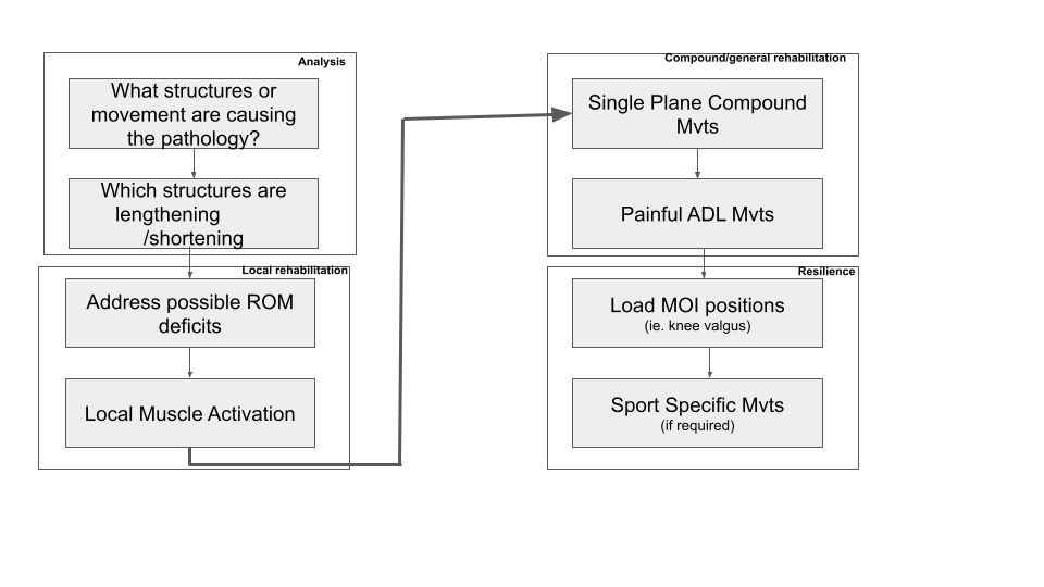
Fig .1 shows the progression from analysis of injury to building resilience Stage 1: Analysis of the injury, ascertaining the mechanism of injury, what structures in the kinetic chain are causing the pathology and what structures are lengthening and shortening.
Stage 2: local structures and muscles, addressing deficits in ROM throughout the kinetic chain and particular muscles that may need additional activation ie VMO activation.
Stage 3: Consists of moving onto the compound or general exercises, then more specific movements that contribute to the main aggravating factors of their pain, ie stair descending.
Stage 4: Building resilience to prevent reinjury, the patient or athlete will need to subject their newly healed structures to the stressors that caused the failure originally
There is a distinct structure of this rehabilitation model. After Stage 1 analysis, outcome measures should be used depending on what is required for the patient’s needs. Such as exercise/activity needs, specific strength requirements etc. If this model is treated more like a flow chart the patient cannot move on to the next stage until the outcome measure is met. Maxime Dougados,(2004) discusses that patient progress should be progressed if suitable improvement has been established either by a reduction in pain levels or similar. Therefore the therapist may consider not progressing the patient or athlete as they haven’t earned the right to move onto Stage 3: Compound/general rehabilitation..
On the other hand, such linear models should be interpreted in shades of grey(Brandt & Pope, 1997), walking for someone might not cause pain but the stairs will. The therapist should not be deterring the patient from compound movements that do not exacerbate their condition.
To conclude, the idea of standardised rehabilitation definitely depends upon the discretion of the therapist establishing set outcome measures before progressing the patient. Rehabilitation is never linear, for some, a generalised type of rehab in a pain-free to mild pain range may be more useful than specialised rehabilitation. Sometimes it will depend on the mechanism of injury, the pathology coupled with the needs and expectations of the person.
References:
Alves Fsm, Oliveira Fs, Junqueira Chbf, Azevedo Bms, & Dionísio Vc. (2009). Análise do padrão eletromiográfico durante os agachamentos padrão e declinado. Brazilian Journal of Physical Therapy, 13(2), 164–172. https://doi.org/10.1590/s1413-35552009005000021
Brandt, E. N., & Pope, A. M. (1997). Models of Disability and Rehabilitation. Nih.gov; National Academies Press (US). https://www.ncbi.nlm.nih.gov/books/NBK233576/
De Baets, L., Meulders, A., Van Damme, S., Caneiro, J., & Matheve, T. (2023). Understanding discrepancies in a person’s fear of movement and avoidance behaviour: a guide for musculoskeletal rehabilitation clinicians who support people with chronic musculoskeletal pain. Journal of Orthopaedic & Sports Physical Therapy, 1–29. https://doi.org/10.2519/jospt.2023.11420
Déborah Viviane Ferrari, Ronaldo Valdir Briani, Danilo, Marcella Ferraz Pazzinatto, Amanda Schenatto Ferreira, Alves, N., & de, M. (2018). Higher pain level and lower functional capacity are associated with the number of altered kinematics in women with patellofemoral pain. Gait & Posture, 60, 268–272. https://doi.org/10.1016/j.gaitpost.2017.07.034
Dhillon, H., Dhillon, S., & Mandeep Singh Dhillon. (2017). Current Concepts in Sports Injury Rehabilitation. Indian Journal of Orthopaedics, 51(5), 529–536. https://doi.org/10.4103/ortho.ijortho_226_17
Gryzlo, S. M., Patek, R. M., Pink, M., & Perry, J. (1994). Electromyographic Analysis of Knee Rehabilitation Exercises. Journal of Orthopaedic & Sports Physical Therapy, 20(1), 36–43. https://doi.org/10.2519/jospt.1994.20.1.36
Hunt, C., Paez, A., & Folmar, E. (2017). THE IMPACT OF ATTENTIONAL FOCUS ON THE TREATMENT OF MUSCULOSKELETAL AND MOVEMENT DISORDERS. International Journal of Sports Physical Therapy, 12(6), 901–907. https://www.ncbi.nlm.nih.gov/pmc/articles/PMC5675366/
Logan, C. A., O’Brien, L. T., & LaPrade, R. F. (2016). POST OPERATIVE REHABILITATION OF GRADE III MEDIAL COLLATERAL LIGAMENT INJURIES: EVIDENCE BASED REHABILITATION AND RETURN TO PLAY. International Journal of Sports Physical Therapy, 11(7), 1177–1190. https://www.ncbi.nlm.nih.gov/pmc/articles/PMC5159640/
Petersen, W., Ellermann, A., Gösele-Koppenburg, A., Best, R., Ingo Volker Rembitzki, Gerd Peter Brüggemann, & Liebau, C. (2013). Patellofemoral pain syndrome. Knee Surgery, Sports Traumatology, Arthroscopy, 22(10), 2264–2274. https://doi.org/10.1007/s00167-013-2759-6
Roush, M. B., Sevier, T. L., Wilson, J. K., D. McEwan Jenkinson, Helfst, R. H., Gehlsen, G. M., & Basey, A. L. (2000). Anterior Knee Pain: A Clinical Comparison of Rehabilitation Methods. Clinical Journal of Sport Medicine, 10(1), 22–28. https://doi.org/10.1097/00042752-200001000-00005
Vilella, R. C., & Anil. (2022, September 5). Musculoskeletal Examination. Nih.gov; StatPearls Publishing. https://www.ncbi.nlm.nih.gov/books/NBK551505/